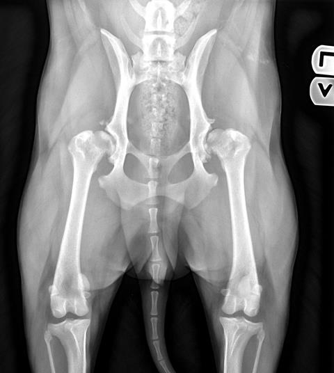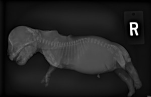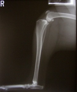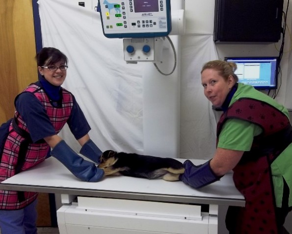
Radiography is a commonly used diagnostic tool in veterinary practice. A fundamental understanding of how radiographs are created enables the user to select the most appropriate exposure factor settings during radiograph production, in order to achieve optimal image quality. Our practice has Direct Digital Radiology for both our regular large whole body x-rays and for the smaller dental xrays. The advantages of digital radiography in a veterinary care facility are widely recognized. We are able to obtain much greater resolution and contrast in our x-rays. Plus the software programs allow us to manipulate the x-rays and get much greater diagnostic value from them. Digital technology uses much less radiation so it is safer for our staff and your beloved pets. We get much higher quality images much faster allowing for quicker and better diagnoses and more efficient treatments. Telemedicine allows us to send digital radiographs or dicom x-rays instantly to board certified radiologists for consultation and interpretation.
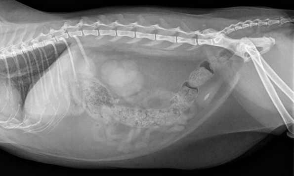
Radiology (x-rays) is routinely used to provide valuable information about a pet’s bones, gastrointestinal tract (stomach, intestines, colon), respiratory tract (lungs), heart, and genitourinary system (bladder, prostate). It can be used alone or in conjunction with other diagnostic tools to provide a list of possible causes for a pet’s condition, identify the exact cause of a problem or rule out possible problems.
When a pet is being radiographed, an x-ray beam passes through its body and hits a piece of radiographic film. Images on the film appear as various shades of gray and reflect the anatomy of the animal. Bones, which absorb more x-rays, appear as light gray structures. Soft tissues, such as the absorb fewer x-rays and appear as dark gray structures. Interpretation of radiographs requires great skill on the part of the veterinarian.

