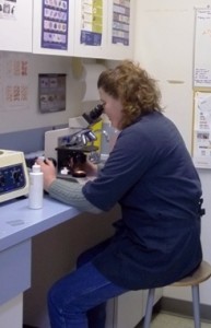
Several types of potential health problems can be identified with laboratory screening: diabetes, liver disease, kidney disease, thyroid disorders, urinary tract infections, etc.—and the earlier
they are found, the better we can manage the disease. We often use simple blood or urine tests to determine if there is any indication or evidence of  underlying diseases in your pets. The results of laboratory tests on a patient are compared to reference ranges established by measuring the laboratory parameters in a group of normal animals. The reference ranges for each laboratory test differ between laboratories and across species. You must always be careful interpreting laboratory tests. An occasional animal will have a value for a laboratory test that falls outside the “normal” reference range, but the value may still be normal for that particular animal.
All of our regular routine blood testing and presurgical screening is done right in clinc so we get rapid accurate results when we need them. Our veterinarians will always interpret the results of laboratory tests in light of the entire clinical evaluation of your pet. Sometimes laboratory tests need to be repeated to evaluate trends, which may provide more information than measurement of a single test.
The results of laboratory tests may be influenced by drugs your pet is receiving and some are influenced by a recent meal. Always provide your veterinarian with information about any drug your pet is receiving. Inquire when you make an appointment for veterinary care, whether you should fast your pet before the visit in case laboratory samples are collected. Additionally, even a completely normal Pet Wellness Report laboratory screening has tremendous value. It gives you peace of mind, and also provides a good baseline so that if the laboratory values begin to change in the future, we will be aware of any trends early–which helps make diseases more manageable and easier to treat.
We have the full line of IDEXX VetLab In-house Analyzers to provide you with accurate real-time service and much faster turn around results so we can start treating your loved one right away.
For some specific tests and complete diagnostic laboratory work up we will send them to the IDEXX lab in Edmonton which takes a few days but often provides greater in depth information.

Below we have provided a quick description of the most common tests we do. Click on this link for more detailed information on what a Complete Blood Count is all about.
Complete blood count (CBC)
The complete blood count measures the number of cells of different types circulating in the bloodstream. There are three major types of blood cells in circulation; red blood cells (RBC), white blood cells (WBC), and platelets. Red blood cells are produced in the bone marrow, which is the soft center of bones. RBCs pick up oxygen brought into the body by the lungs, and bring that oxygen to cells throughout the body. Red blood cells live in the blood stream for about 100 days although the actual time varies with the type of animal. Old red blood cells are removed from the blood stream by the spleen and liver. Red blood cell numbers can be decreased (anemia) if they are not produced in adequate numbers by the bone marrow, if their life span is shortened (a condition called hemolysis), or if they are lost due to bleeding. Increased red blood cell numbers is called polycythemia and is usually due to concentration of the blood due to dehydration.
The complete blood count also includes a measure of hemoglobin, which is the actual substance in the red blood cell that carries oxygen.
There are several types of white blood cells in blood, including neutrophils (PMNs), lymphocytes, monocytes, eosinophils and basophils. Lymphocytes are produced in lymph nodes throughout the body. The other white blood cell types are produced in the bone marrow along with the red blood cells and platelets. The majority of white blood cells in circulation are neutrophils, which help the animal fight infections. Neutrophils can be decreased in pets with bone marrow disease, in some viral diseases, and in some pets receiving cancer chemotherapy drugs. Neutrophils are increased in pets with inflammation or infection of any part of the body and in pets receiving prednisone or other cortisone-type drugs.
Lymphocytes also help fight infection and produce antibodies against infectious agents (viruses, bacteria, etc.). Lymphocytes may be increased in puppies and kittens with an infection, they can be decreased in pets who are severely stressed, and lymphocytes might be lost in some types of diarrhea. Certain drugs, such as prednisone (a cortisone-type drug) will decrease the number of lymphocytes in the blood stream.
Monocytes may be increased in pets with chronic infections. Eosinophils and basophils are increased in pets with allergic diseases, or parasitic infections (worms, fleas, etc.).
Platelets are produced in the bone marrow and are involved in the process of making a blood clot. Platelets live a few weeks and are constantly being produced by the bone marrow. Low platelet counts occur if the bone marrow is damaged and doesn’t produce them, or if the platelets are destroyed at a faster rate than normal. The two primary causes of platelet destruction are immune-mediated destruction (ITP or IMT) and DIC (disseminated intravascular coagulation). Immune-mediated thrombocytopenia happens when the animal’s immune system destroys platelets. DIC is a complex problem in which blood clots form in the body using the platelets faster than the bone marrow can produce new ones. Animals with a low platelet count bruise easily and may have blood in their urine or stool.
Packed cell volume (PCV) (called hematocrit, HCT, in humans) is another measure of red blood cells. A small amount of blood is placed in a tiny glass tube and spun in a centrifuge. The blood cells pack to the bottom of the tube and the fluid floats on top. The PCV is the percent of blood, that is cells, compared to the total volume of blood. In normal dogs and cats, 40-50% of the blood is made up of blood cells and the remainder is fluid.
Blood and urine tests are performed to get an initial overview of the health, and sometimes the function, of body organs. Some blood tests are very specific for a single organ, whereas other tests are affected by several organs. Blood tests are often performed as a biochemistry profile, or chemistry panel, which is a collection of blood tests to screen several organs at one time. The makeup of a biochemical profile varies with the laboratory in which it is performed. Following are some of the more commonly performed chemical tests:
Albumin (ALB) is a small protein produced by the liver. Albumin acts as a sponge to hold water in the blood vessels. When blood albumin is decreased, the pressure created by the heart forcing blood through the blood vessels causes fluid to leak out of the blood vessels and accumulate in body cavities such as the abdominal cavity or in tissues as edema. Albumin is decreased if the liver is damaged and cannot produce an adequate amount of albumin or if albumin is lost through damaged intestine or in the urine due to kidney disease. The only cause of increased albumin is dehydration.
Alkaline phosphatase (ALKP) originates from many tissues in the body. When alkaline phosphatase is increased in the bloodstream of a dog the most common causes are liver disease, bone disease or increased blood cortisol either because prednisone or similar drug is being given to the pet or because the animal has Cushing’s disease (hyperadrenocorticism). In cats, the most common causes of increased alkaline phosphatase are liver and bone disease.
Alaninine Aminotransferase (ALT) is an enzyme produced by liver cells. Liver damage causes ALT to increase in the bloodstream. ALT elevation does not provide information as to whether the liver disease is reversible or not.
Amylase (AMYL) is an enzyme produced by the pancreas and the intestinal tract. Amylase helps the body breakdown sugars. Amylase may be increased in the blood in animals with inflammation (pancreatitis) or cancer of the pancreas. Sometimes pancreatitis is difficult to diagnose and some dogs and cats with pancreatitis will have normal amounts of amylase in the blood. Lipase is another pancreatic enzyme which is responsible for the breakdown of fats and which may be increased in patients with pancreatic inflammation or cancer.
Bile acids are produced by the liver and are involved in fat breakdown. A bile acid test is used to evaluate the function of the liver and the blood flow to the liver. Patients with abnormal blood flow to the liver, a condition known as portosystemic shunt will have abnormal levels of bile acids. The bile acid test measures a fasting blood sample and a blood sample two hours after eating.
Bilirubin (BIL) is produced by the liver from old red blood cells. Bilirubin is further broken down and eliminated in both the urine and stool. Bilirubin is increased in the blood in patients with some types of liver disease, gallbladder disease or in patients who are destroying the red blood cells at a faster than normal rate (hemolysis). Large amounts of bilirubin in the bloodstream will give a yellow color to non-furred parts of the body, which is called icterus or jaundice. Icterus is most easily recognized in the tissues around the eye, inside the ears and on the gums.
BUN (blood urea nitrogen) is influenced by the liver, kidneys, and by dehydration. Blood urea nitrogen is a waste product produced by the liver from proteins from the diet, and is eliminated from the body by the kidneys. A low BUN can be seen with liver disease and an increased BUN is seen in pets with kidney disease. The kidneys must be damaged to the point that 75% of the kidneys are nonfunctional before BUN will increase. Pets that are severely dehydrated will have an increased BUN as the kidneys of a dehydrated patient don’t get a normal amount of blood presented to them, so the waste products do not get to the kidneys to be eliminated.
Calcium (Ca) in the bloodstream originates from the bones. The body has hormones, which cause bone to release calcium into the blood and to remove calcium from the blood and place it back into bone. Abnormally high calcium in the blood occurs much more commonly than low calcium. High blood calcium is most commonly associated with cancer. Less common causes of elevated calcium are chronic kidney failure, primary hyperparathyroidism which is over-function of the parathyroid gland, poisoning with certain types of rodent bait and bone disease.
Low blood calcium may occur in dogs and cats just before giving birth or while they are nursing their young. This is called eclampsia and occurs more commonly in small breed dogs. Eclampsia causes the animal to have rigid muscles which is called tetany. Another cause of low blood calcium is malfunction of the parathyroid glands which produce a hormone (PTH) that controls blood calcium levels. Animals poisoned with antifreeze may have a very low blood calcium.
Cholesterol (CHOL) is a form of fat. Cholesterol can be increased in the bloodstream for many reasons in dogs. It is much less common for cats to have increased cholesterol. Some of the diseases that cause elevated cholesterol are hypothyroidism, Cushing’s disease, diabetes and kidney diseases that cause protein to be lost in the urine. High cholesterol does not predispose dogs and cats to heart and blood vessel disease as it does in people.
Creatinine (CREA) is a waste product that originates from muscles and is eliminated from the body by the kidneys. An elevation of creatinine is due to kidney disease or dehydration. Both creatinine and BUN increase in the bloodstream at the same time in patients with kidney disease.
Creatinine kinase (CK) is released into the blood from damaged muscle. Elevation of creatinine kinase therefore suggests damage to muscle including heart muscle.
Glucose (GLU) is blood sugar. Glucose is increased in dogs and cats with diabetes mellitus. It may be mildly increased in dogs with Cushing’s disease. Glucose can temporarily increase in the blood if the dog or cat is excited by having a blood sample drawn. This is especially true of cats. A quick test to determine whether a glucose elevation is transient or permanent is to look at the urine. If the glucose is chronically elevated there will be an increased amount of glucose in the urine as well.
Low blood sugar occurs less commonly and can be a sign of pancreatic cancer or overwhelming infection (sepsis). Low blood sugar can cause depression or seizures. Low blood sugar can be seen if the blood sample is improperly handled. Red blood cells will use glucose so typically red blood cells are removed from the blood sample and the clear part of the blood (plasma or serum), is used for analysis.
Phosphorus (PHOS) in the bloodstream originates from bones and is controlled by the same hormone, PTH (parathyroid hormone) which controls blood calcium. Phosphorus is increased in the bloodstream in patients with chronic kidney disease. Like BUN and creatinine, phosphorus increases in these patients when about 75 percent of both kidneys is damaged.
Potassium (K) is increased in the bloodstream in the pet with acute kidney failure such as kidney failure caused by antifreeze poisoning, in dogs with Addison’s disease and in animals with a ruptured or obstructed bladder.
Potassium is lost from the body in vomit, diarrhea and urine. Pets that are not eating may have a low blood potassium. Low blood potassium can cause the pet to feel weak. Cats with low potassium may develop painful muscles.
Sodium (Na) may be slightly increased in the blood if the patient is dehydrated although many dehydrated dogs and cats have a normal blood sodium. Low blood sodium is most commonly seen with Addison’s disease (hypoadrenocorticism).
Total protein (TP) includes albumin and larger proteins called globulins. Included in the globulins are antibodies which are protein molecules. Total protein can be increased if the dog or cat is dehydrated or if the pet’s immune system is being stimulated to produce large amounts of antibody. Total protein is decreased in the same situations which reduce albumin or if the pet has an abnormal immune system and cannot produce antibodies.
Urinalysis (UA): A urine sample can provide information about several organ systems. The concentration, color, clarity and microscopic examination of the urine sample can provide diagnostic information.
Urine may be obtained by catching a sample during normal urination, by passing a catheter into the bladder or by placing a small needle through the body wall into the bladder, a procedure called cystocentesis. Depending upon why the urine sample is being collected, one collection method may be preferred over another. Inquire at the time you make an appointment for veterinary care if a urine sample may be collected. Preventing your pet from urinating prior to the appointment will assure that your pet’s bladder will contain urine for sampling.
Adapted and modified from Washington State University which assumes no liability for injury to you or your pet incurred by following these descriptions or procedures.

